St Andrews Biophotonics Summer School 2024
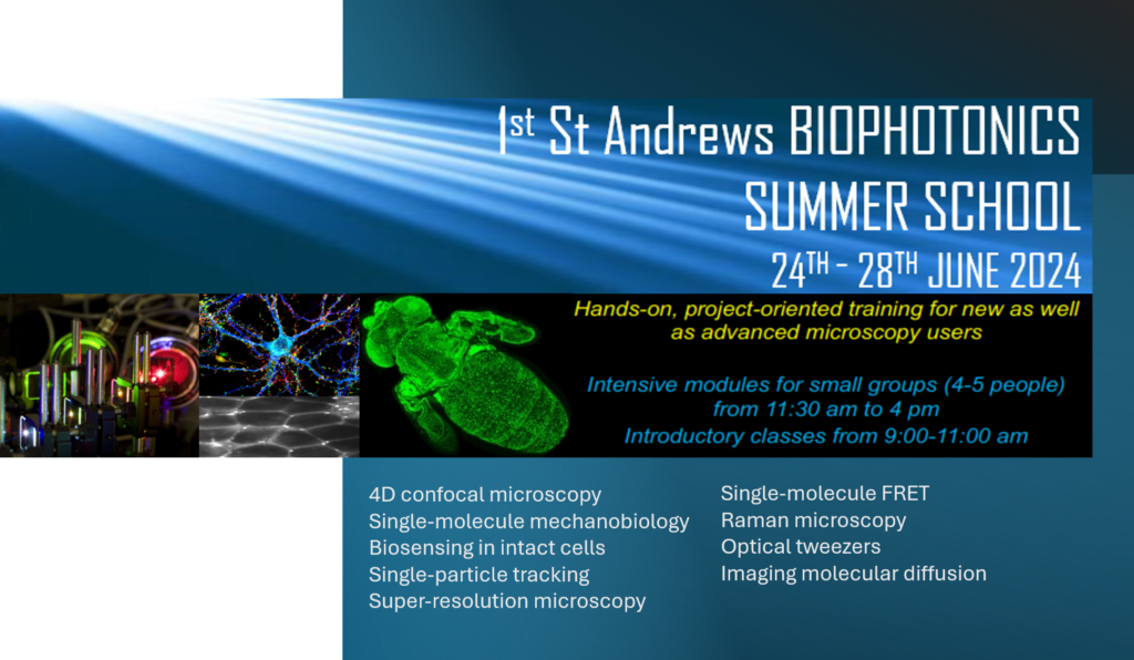
The Centre of Biophotonics (CoB) at st Andrews is launching its 1st Summer School in Biophotonics. The school is essentially for PhD students, post-docs and early career scientists working across the Physics-Life Science Interface. The aim of the School is to provide participants with a solid background in cutting-edge microscopy techniques including single-molecule fluorescence, super-resolution, tracking, single-molecule force detection using optical tweezers, 4D confocal microscopy and Raman sensing. Specific details about these topics are outline in sections below.
The school is running mostly in a hands-on format with a short introduction to the microscopy principles of each project-oriented exercise during the mornings from 9.00 to 11.00. This will be followed by experimental case studies on our facilities at the CoB in small groups of 4-5 people. These hands-on project and the morning lecture will be delivered by group leader experts on each respective area.
The summer school has a limited number of spaces: 40.
Contact information:
Prof Carlos Penedo. Director Centre of Biophotonics. [email protected]
Dr Paolo Annibale, School of Physics and Astronomy, [email protected]
Registration
The registration is kept low (£150 internal St Andrews University participants, £270 for external attendes) and includes coffee breaks and lunch every day.
To register click here
Accomodation
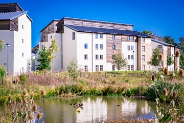
En suite B&B accommodation at David Russell apartments at a discounted rate for the nights of Sunday 23rd June to Thursday 27th June inclusive using the promotional code SBSS24 can be booked separately. The online accomodation booking system will be available until the 24th April 2024, please book before that deadline here
Topics
Topic 1: Confocal microscopy basics and in vivo imaging of Drosophila pupae using 4D confocal laser-scanning microscopes. Instructor: Dr. Marcus Bischoff
This module will discuss fundamentals of confocal imaging applied to in vivo imaging of Drosophila pupae expressing GFP-tagged proteins. We will dissect and mount pupae for microscopy. We will then image them using a Leica SP8 confocal microscope. Concepts that will be explored are image saturation, bleed-through and spectral unmixing, z-sectioning, time-series imaging, pitfalls of in vivo microscopy, programming macros for automated imaging, tiling, FRAP, resonant scanning & galvo stage. We will also use a Zeiss Airyscan confocal to image pupae in moderate super-resolution.
Topic 2: Single-molecule mechanobiology using confocal-force microscopy. Instructor: Prof Carlos Penedo
Dual-trap optical tweezers combined with confocal fluorescence. In this module we will go through the basis of single-molecule force applications to study biomolecules and their combination with confocal fluorescence. We will perform experiments to study the mechanical stress of the DNA double helix using force-extension curves whilst visualizing its melted state using fluorescent intercalators. We will also explore the binding of a protein to the DNA structure as a function of applied force. These experiments will be carried out in a LUMICKS C-trap Dymo microscope.
Topic 3: Single molecule structural dynamics by FRET. Instructors: Dr. Alfonso Brenlla and Prof Carlos Penedo
In this module we will explore the use of PRISM-based total-internal excitation to visualize individual molecules labelled with a FRET pair. We will carried out experiments to visualize DNA conformational changes and protein-DNA interactions. We will explain the basis of the optical setup, immobilization strategies the range of data that can be obtained for structural dynamics using MATLAB-based software.
Topic 4: Molecular diffusion by spatio-temporal correlations. Instructors: Dr. Paolo Annibale, Jothi-Letchoumi Kumar
This module will explore how to measure the diffusion of fluorescently tagged intracellular proteins using spatial-temporal image correlations. Specifically, rapid linescans will be collected in a Leica SP8 microscope and diffusion onformation recover from spatial-temporal correlations
Topic 5: Biosensing in intact cells using FRET. Instructors: Dr. Paolo Annibale, Debasmita Banik and Soumyabrata Banik
FRET biosensing module: In this module we will explore how Fluorescence Resonance Energy Transfer can be applied to measuring intracellular second messenger in isolated cells. We will conduct sensitized emission measurements in an epifluorescence microscope, using a beamsplitter to monitor donor and acceptor signals on an EM-CCD camera. We will be using a ratiometric biosensor, widely reported in the literature. We will be using a setup comprising an environmental chamber and will monitor the effects of different ligands on the FRET response. Practical aspects concerning data interpretation and analysis will be discussed in detail.
Topic 6: Molecular diffusion by single-particle tracking. Instructors: Dr. Juan Varela, Dr Vanya Metodieva, Chloe O’Rourke
This module will explore how to measure the diffusion of membrane receptors tagged with quantum dots in living cells. The session will explore the aspects of receptor labelling, trajectory reconstruction and measurement of mean-squared displacements and diffusion coefficients.
Topic 7: Non-intrusive detection of molecular fingerprints using Raman. Instructor: Dr. Graham Bruce
This module will explore the fundamentals and practical steps of acquiring Raman spectra for molecular profiling of samples, including samples hidden behind barriers. The technique provides a non-destructive and non-contact detection method for precise analysis of chemical composition, and examples will include alcohols in sealed bottles.
Topic 8: Imaging the nanoscale organization of protein machinery using super-resolution microscopy. Instructor: Dr John Danial
In this module we will discuss the fundamentals of Super Resolution Microscopy (SRM) – microscopy methods that break the diffraction limit of light to image biological specimen with nanometre resolution. We will explore the different classes of SRMs, including Single Molecule Localisation Microscopies (SMLMs) such as Photo Activated Localisation Microscopy (PALM), Stochastic Optical Reconstruction Microscopy (STORM), and Points Accumulation in Nanoscale Topography (PAINT); position targeted microscopies, such as Stimulated Emission by Depletion (STED); as well as state-of-the-art methods that aim to push precisions down to the Angstrom scale. Towards the end, we will use a custom-built microscope to image nanorulers made out of DNA to practically demonstrate how SRM works.
Summer school programme timetable
Coming soon