Introduction
Introduction to the CoB
Research efforts at the CoB cover all stages of biological function and human disease, from gaining fundamental understanding of the complex underlying mechanism to the development of early-detection diagnostic markers and sensing methods, and from volumetric imaging across tissue using specifically tailored microscopes to the use of light to mechanically manipulate individual proteins, DNA, RNA and cells and tissues.
Importantly, the CoB is also strongly committed to training the next generation of scientist working at the interface between disciplines. Our vision is to facilitate a training environment where students are encouraged to think more fundamentally about light-based technologies, to identify new challenges and opportunities, and by doing so, innovate and drive entrepreneurial projects forward in their future careers whether in industry, academia, humanities or elsewhere.
Early days of biophotonics at St Andrews
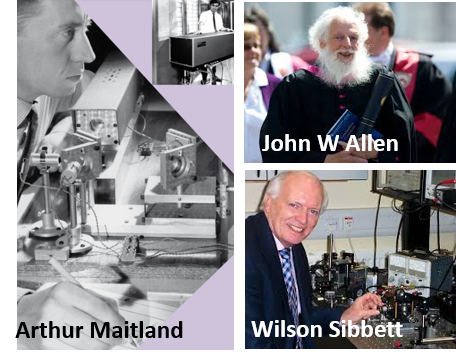
St Andrews University has a strong track-record in Photonics innovation that dates back to early 1960s when Arthur Maitland started the laser physics group in 1963 (Left picture). Maitland’s research into gas discharge lasers led to the development of a gold metal vapour laser in the late 1980s. The laser was used in photodynamic therapy (PDT) treatment by clinicins at Ninewells Hospital in Dundee, thus becoming the first of a long list of collaborations between both institutions, that remains to the present days.
Further advances in photonics continued at St Andrews for the next decades including the pioneering work by John W. Allen (Right top picture) and Wilson Sibbett (Right bottom picture). John W Allen developed the world’s first practical, visible light-emitting diode (or LED as we know it). In 1972 he became the founding Director of the Wolfson Institute of Luminescence and in 1980 was awarded a Personal Chair in Solid-State Physics. In 1989 Sibbett’s group developed the technique of Kerr-lens mode-locking and opened the floodgates to the field of practical ultra-fast spectroscopy and its application in Biology. Biophotonics in St Andrews received a significant footing from grants in 2004 consisting of an EPSRC Bioplatform grant and a CRUK interdisciplinary grant. The latter brought together researchers from three different Schools: Physics (Profs Dholakia/Samuel/Krauss), Biology (Profs Hay/Gunn-Moore) and Medicine (Profs Riches/Bryant/Herrington). Since then, Biophotonics research has attracted more than £10M in external funding and it has been recognized as a strategic research area within the University of St Andrews.
Vision for the future
Biophotonics has come a long way over the past few decades and advances in light-based technologies have resulted in transformative tools to study and manipulate biological objects with an unprecedented level of detail. The area is expected to play a key role in shaping the next generation of diagnostic, sensing tools and imaging modalities of the 21st century.
By integrating teams of molecular and cellular biologists, photonics experts, computational scientists and technique builders, the CoB is uniquely positioned to contribute towards this goal. In the scheme on the right, we summarized the mid-to long-term challenges addressed by the CoB. These include the implementation of imaging techniques from single molecules to tissues and whole organisms, the integration of imaging and mechanical manipulation methods at molecular and cellular level and the improvement of large dataset analysis by using ‘big data’ and ‘machine learning’ strategies.
Nano- to microscale imaging
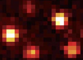
The origins of disease and biological function are found at the molecular level. Advances in single-molecule imaging methods below the diffraction limit are required to capture structural changes and dynamic events underlying the function of molecular machines crucial to cell survival. Specifically, the CoB aims to push the limits in the following areas:
- Development of brighter and more specific probes to increase single-molecule signal-to-noise ratio.
- Unveil molecular mechanisms at smaller and smaller size scales.
- Advance in data analysis protocols to extract rare signals from biological noise.
Image courtesy of Carlos Penedo, Sci. Adv. 4, eaao5786 (2020)
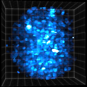
Meso- to macroscale imaging
In vivo volumetric imaging of small model organisms such as C. elegans, Drosophila, Zebrafish, Amphioxus and larger animals such as rodents has substantially advanced in recent years but challenges still remain in the following areas:
- Improve our understanding of light-beam shaping and light-scattering to enhance signal quality.
- Develop better 3D rapid scanning techniques of large volumes.
- Protocols to correlate images from the same of different techniques and data across large spatial scales from nanometers to millimetres and beyond.
(Image courtesy of Maarten Zwart)
Multi-modal methods
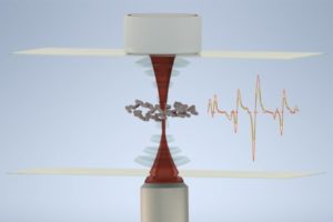
Understanding the complexity of biological systems will greatly benefit from the ability to record in parallel information extracted from different techniques. Moreover, there is a growing need to combine manipulation/immobilization and imaging methods to move actively alter the state of the system in real time. At the CoB, we aim to develop hybrid methods combining acoustic, optical and magnetic traps with fluorescence imaging and Raman spectroscopy, super-resolution and electron-microscopy or fluorescence detection whilst applying mechanical stress using optical or magnetic tweezers.
Image courtesy of Kishan Dholakia, Commun. Bio. 3, 235 (2020)
Computational modelling/image processing
As in many other research areas, the size of the datasets generated in biophotonics are continuously becoming larger and increasing in complexity. Currently, we only scratch the surface of the potential information contained in these imaging datasets. At the CoB, we integrate instrument developers with computational experts and light-propagation modellers to enhance the ability to unmask hidden information and correlate information extracted from the same sample by different techniques.
Image courtesy of Kishan Dholakia, Sci. Rep. 10, 8090 (2020)
Nanophotonics
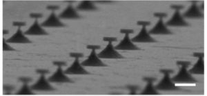
Miniaturization of lasers for intracellular function, multiplexed detection and imaging, and the application of nanoparticles for sensing, targeted delivered, plasmonics and surface-enhanced Raman scattering are just some examples of recent advances where the convergence of nano-patterning and biophotonics is increasing the ability to shape the properties of light at the nanoscale.
Image courtesy of Andrea di Falco, Malte Gather, Marcel Schubert and Simon Powis, Nat. Comm.9, Article number: 4817 (2018)
Education and training
Biophotonics is intrinsically an inter-disciplinary subject with elements from many disciplines and requires scientists with broad knowledge in diverse areas including biology, optics, and image processing. At the CoB, we are committed to educating and training the next generation of researchers and we are continuously working in the development of innovative education programs at all levels, from undergraduate to early-career scientists. Our goal is to prepare students and researchers with specific training so that they can apply and further develop optical methods in bioscience research both in academia and industry.