Gallery
Below we show representative images and videos as examples of the range of techniques, scales, and samples investigated within the CoB.
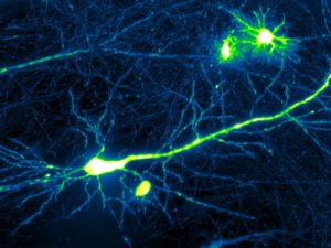
Calcium flux in a living mouse brain section expressing a genetically encoded calcium indicator. Stimulation was performed using a pulse of hyperkalemic solution. [Tello lab]
Contractility mapping of the zebrafish heart using light-sheet microscopy with acoustic sample confinement. Scale bar is 50 μm. Yang et al. Nature Communications 10, 669 2020. [Somorjai lab] [Dholakia lab]
Two larval epithelial cells undergo pulsed contractions during Drosophila abdominal morphogenesis. The cells’ actin cytoskeleton is labelled with a GFP-reporter. Pulido-Companys et al. J. Cell Sci., 133 (6) 2020. [Bischoff lab]
Large field of view fluorescence video-rate microscopy of neonatal cardiomyocytes . Cells were labelled with SiR-actin to visualize sarcomeric actin. [Schubert lab] [Pitt lab] [Miles lab] [Gather lab]
Drosophila abdominal morphogenesis. Histoblasts replace larval epithelial cells during formation of the adult epidermis. Nuclei of both cell types are labelled with Histone-GFP. Bischoff and Cseresnyes, Development 136, 2403 (2009). [Bischoff lab]
DIC microscopy time-lapse video of a spontaneously beating neonatal cardiomyocyte showing an internalized microlaser as circular object in the centre of the cell. Schubert et al, Nat. Photonics, 15, 452, 2020. [Schubert lab] [Pitt lab] [Miles lab] [Gather lab]
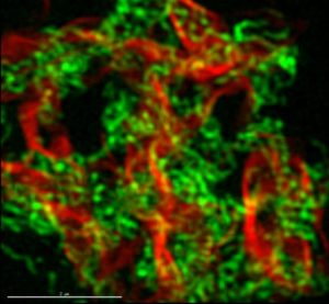
Drosophila abdominal morphogenesis. Histoblasts replace larval epithelial cells during formation of the adult epidermis. Nuclei of both cell types are labelled with Histone-GFP. In addition, cells of the P compartment are labelled with a nuclear red fluorescent marker (Bischoff and Cseresnyes, Development 136, 2403 (2009). [Bischoff lab]
Neural circuits imaged using a planar Airy light-sheet microscopy. [Vettenburg lab]
Amphioxus head as seen under a fluorescence microscope. [Somorjai lab]

Widefield two-photon fluorescence image of a smiley face using temporally focussed illumination (represented in faux colour) Escobet-Montalban et al . 2020, Vol. 4, no. 10, eaau1338 [Dholakia lab][Mazilu lab]
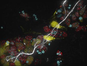
Directed differentiation of mouse embryonic stem cells into neurons. Sox1 marker of neural progenitors in yellow, neurofilaments in white, splicing speckles in red and DNA in cyan. [Sleeman lab]
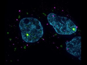
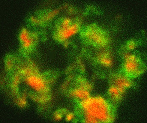
Cytoplasmic stress granules (green) and P-bodies (magenta) in a model of Myotonic Dystrophy Type 1 (nuclei shown in Cyan). [Sleeman lab]
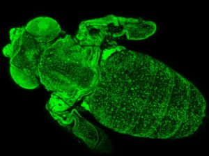
Drosophila pupa removed from its pupal case. All cells are labelled with Histone-GFP. [Bischoff lab]
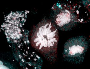 Mouse embryonic stem cells undergoing cell division. DNA chromosomes) in white. [Sleeman lab]
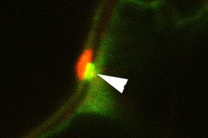
Potato virus X capsid protein (green) inserted into plasmodesmata, triple gene block (TGB) 3 movement protein accumulating outside. Tilsner et al. J. Cell Biol. 201, 981 (2013). [Tilsner lab]
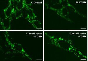
Direct 2NBDG assay-leptin increases neuronal glucose uptake. CGSD (combined glucose and serum deprivation). Scale bar: 10 micrometers. [Doherty lab]
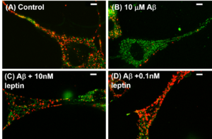
JC-1 assay-leptin improves membrane potential induced by amyloid beta. Scale bar: 10 micrometers. [Doherty lab]

Directed differentiation of mouse embryonic stem cells into neurons. Sox1 marker of neural progenitors in yellow, neurofilaments in white, splicing speckles in red and DNA in cyan. [Sleeman lab]
Potato virus X replication ‘factory’. Triple gene block (TGB) 1 movement protein aggregates (red) surrounded by viral RNA (green) labelled with Pumilio-BiFC. Tilsner et al. Plant Phys. doi: 10.1104/ pp.111.189605 (2012) [Tilsner lab]

Fluorescent imaging of muscle activation during locomotion in Drosophila larvae using green fluorescent protein. Red: image sequence showing peristaltic wave of muscle contraction corresponding to forward locomotion, Blue: peristaltic wave during backward locomotion, Green: muscle activation during head sweep behaviour. [Pulver lab]
Light-sheet microscopy of cell and tissue behaviours during primitive streak formation in Myr-GFP embryos. Robzicki et al. Nat. Cell Biol. 17, 397, 2015 [MacDonald lab]

2 cm by 2 cm OLEDs on flexible plastic substrate designed for antimicrobial photodynamic therapy (PDT). OLEDs are promising light sources for PDT as they are lightweight, ultrathin and uniform. They have potential to be flexible and disposable to suits the needs for ambulatory treatment.
2 cm by 2 cm OLEDs on flexible plastic substrate designed for antimicrobial photodynamic therapy (PDT). OLEDs are promising light sources for PDT as they are lightweight, ultrathin and uniform. They have potential to be flexible and disposable to suits the needs for ambulatory treatment.
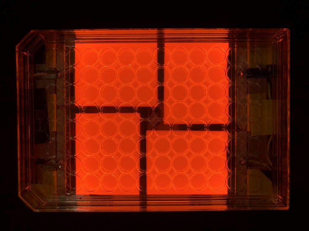
Four large area OLEDs on a glass substrate designed for biological experiments on 96 well-plate. Each of OLED pixel has an individual power switch and it can illuminate 12 wells at the same time. This device provides uniform illumination during the biological experiments and improves the throughput on a single trial.
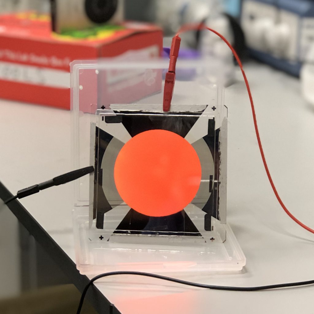
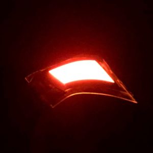
Large area OLED in round shape for potential clinical PDT applications. [Samuel lab] [Turnbull lab]
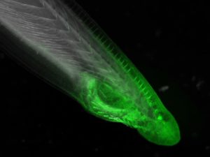
Amphioxus head as seen under a fluorescence microscope. [Somorjai lab]
Widefield two-photon fluorescence image of a smiley face using temporally focussed illumination (represented in faux colour) Escobet-Montalban et al . 2020, Vol. 4, no. 10, eaau1338 [Dholakia lab][Mazilu lab]
Directed differentiation of mouse embryonic stem cells into neurons. Sox1 marker of neural progenitors in yellow, neurofilaments in white, splicing speckles in red and DNA in cyan. [Sleeman lab]
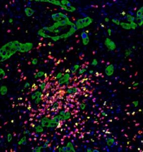
Multiplexed image analysis by HALO software. Colorectal cancer cells (green) and lymphocytes (CD3: yellow & CD8: red). [QUAD lab]
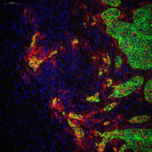
Bladder cancer cells (green) and lymphocytes (blue) heterogeneusly expressing PD-L1 (red). [QUAD lab]
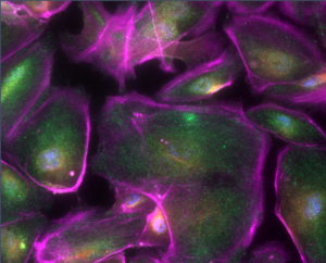
Multiplexed fluorescence imaging of podocytes. Blue: nuclei, Purple: actin, Green: Nrf1, Red: Nq01. [QUAD lab]
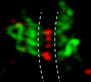
Potato virus X Triple gene block (TGB) 1 movement protein (red) inserted into plasmodesmata, triple gene block (TGB) 2 movement protein (green) accumulating in modified ER tubules outside. 3D-SIM reconstruction, position of cell wall marked by dotted line. Tilsner et al., J. Cell Biol. 201, 981 (2013). [Tilsner lab]
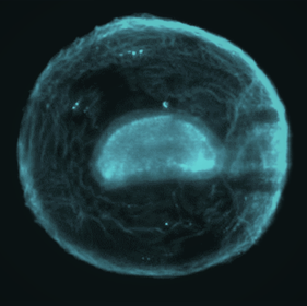
Amphioxus embryo confined in an acoustic trap and imaged using light-sheet microscopy. Yang et al. Nature Communications, 10, Article 669, (2019) [Dholakia lab][Somorjai lab]
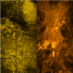
(Left) Surface of a normal colon tissue, (right) surface of a cancerous one. Images taken with the open top light sheet with a resolution of 2 microns. Corsetti et al, OSA Continuum, 3, 1068 (2020) [Dholakia lab]
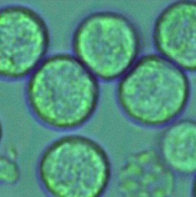
Immune cells imaged with digital holographic microscopy. Gupta et al . Optics Express, 27, 13706 (2019), [Dholakia lab] [Powis lab]
Potato virus X Triple gene block (TGB) 1 movement protein aggregates (red) surrounded by triple gene block (TGB) 2 movement protein (green) accumulating in modified ER tubules. 3D-SIM reconstruction. Linnik et al. Front. Plant Sci. (2013) doi: 10.3389/f pls.2013.00006. [Tilsner lab]